Science in the lab across scales
How are barriers formed and disrupted in the gut? We address this question by working across tissue-scale and organotypic models, using patient-derived organoids and human tissues to investigate how epithelial junctions organize at the nanoscale and shape tissue architecture. We are developing ways to archive higher quantitative information on large and complex volumetric samples using LLM- based approaches for segmentation. By linking junctional organization to morphological changes, we aim to understand how structural disruptions translate into functional defects in diseases such as IBD.
What drives adhesion at cell-cell interfaces? The collective behavior of molecules gives rise to functional complexes that drive cellular organization. We investigate how biomolecules such as proteins, lipids, and glycans dynamically organize at cell–cell interfaces to understand how their coordinated structure leads to function. We are exploring how different self-organization mechanisms including the formation of membrane condensates evolve and regulate cell-cell interactions.
How do biomolecular condensates contribute to disease? We collaborate with diverse teams to investigate how condensates and membraneless compartments drive pathological processes such as ALS.
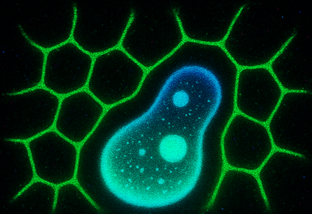
What tools best answer the biology? We leverage cutting-edge technologies to drive discovery, combining STED super-resolution microscopy with adaptive optics to visualize and dynamically track processes at the nanoscale. We further exploit the optical properties of biological samples using approaches such as FLIM, FCS, FRAP, and live-cell imaging to probe molecular behavior and dynamics in their native context.